
- The crown of the dog’s tooth
- The basic forms of crowns in dogs
- The root of the dog’s tooth
- What tooth contains from?
Before talking about Dog’s Teeth Anatomy, let’s define what is dog’s teeth are? Teeth are organs of the oral cavity that perform various functions, the main of which is the mechanical processing of food. The general plan of the structure of the teeth is characteristic for both generations (milk and permanent). Anatomically, a crown, neck, and root are distinguished in a tooth.
Dog’s Teeth Anatomy: The crown of a dog’s tooth
(corona dentis – latin) is the part of it that extends over the gingival margin. The crown represents the main working part of the tooth. There are interdental spaces between the crowns of the teeth, which are covered with gingival papillae. The function of these papillae is to prevent food particles from entering the spaces between the medial and distal surfaces of the tooth crown.
As a result of a number of changes that took place in the process of phylogenesis, in dogs, the crowns of the teeth acquired a different shape – the homodont (same-toothed) dental system became heterodont (multi-toothed). A gradual change in the shape of the tooth and its parts continues at the present time, depending on the changing conditions of detention and various environmental factors.
The basic forms of crowns in dogs
are as follows: spatula (incisors), conical (canines), cylindrical bicuspid (premolars or bicuspidates), and cylindrical multi-cusp (molars). The collum dentis (latin) is the transition point of the crown to the root, hidden under the gingival margin. It connects these two parts of the tooth and is designated as an interception between them. At the neck, the enamel ends, and the enamel (cuticle) is connected to the inner epithelial lining of the gingival margin. Thus, the continuity of the integumentary tissues of the body is created, which serves as an external barrier.
The formation of a tooth neck is associated with the need to separate the working part of the tooth (crown) from the fixing part (root). This makes it possible to increase the physical load on the teeth during vertical chewing and to reduce the traumatic factor on the gums due to a tighter adherence of the mucous membrane to the teeth. The mucous membrane that encloses the teeth is, as it were, under the protection of the teeth with pressure on them directed from the apex of the crown to its base.
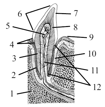
Dog’s Teeth Anatomy: The root of the dog’s tooth
(radix dentis) is the part that is immersed in the alveolus of the jaw. The root is tightly connected to the periosteum (periodontium). The main function of the tooth root is to fix and support the entire tooth. In contrast to the crown and neck, which are single in a tooth, the root is presented in two, three, and more.
The single root is the simplest of the so-called true roots; the appearance of several roots is the result of the complication of the original form of the true root. Inside the crown of the tooth, there is a cavity, which in the roots passes into canals that open at the tops of the roots with holes. The cavity follows the shape of the tooth crown
A tooth contains both soft and hard tissues.
Soft tissues include the pulp, which fills the cavity of the crown and root canals, and the periodontium, which connects the tooth root to the alveoli. Hard tissues of the tooth include enamel, dentin, and cement. The main mass of the tooth in the area of the crown, neck, and roots is dentin, which limits the cavity of the crown of the tooth and root canals. The surface of the dentin of the crown is covered with enamel and the dentin of the root is covered with cement. The complex of supporting tooth tissues (cement, periodontium, bony alveoli, and gums) is called the periodontium.